September 2015 | VOL. 14, NO. 9 | www.McGowan.pitt.edu
Pittsburgh’s 25th Anniversary of First VAD Patient to Be Discharged from a Hospital
Twenty-five years ago a historic medical event occurred in Pittsburgh. In 1990, Brian Williams, a teenager from Georgia, was the first ventricular assist device (VAD) patient to be discharged from the hospital with formal approval from the Food and Drug Administration. Mr. Williams was discharged from the University of Pittsburgh Medical Center’s Presbyterian Hospital to a local Family House, where he lived with his parents while waiting for a heart transplant. As amplified below, substantial progress has been made in the size and reliability of VAD systems since 1990 through collaborative efforts between McGowan faculty and device manufacturers. Over the years, Mr. Williams has told his incredible story to audiences of bioengineers, including students and faculty nationwide. He also has experienced much happiness in his life. He has completed his undergraduate and graduate education, is married to Jenny, and has a son, Jacob.
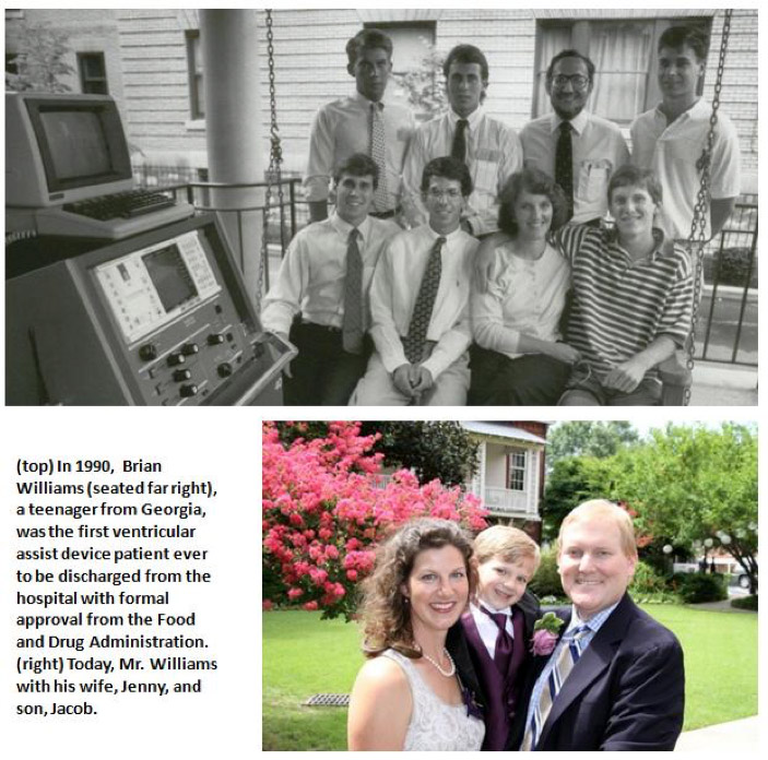
VADs are mechanical devices that help the heart pump blood from one of the main pumping chambers to the rest of the body. VADs have been used since the mid-1980s. In their early days, however, VADs were considered only as a short-term solution for a defective heart. The device was bulky, had a large support console (see console at the left in the 1990 photo above), and required patients to be hospitalized in an intensive care unit. The size of these early VAD pumps excluded most women and children from being candidates for this therapy.
A breakthrough came when McGowan Institute researchers focused on the support of patients with an implantable VAD. Also, these engineers and clinicians were among the first in the world to visualize the VAD as a resource for destination therapy—where the VAD is intended to be the final therapy, vs. serving as a bridge to a transplant for people awaiting a transplant. Today, in certain select cases, VADs can help patients recover from heart failure, making transplantation unnecessary.
“Over the years, Institute clinicians, bioengineers, and students have worked together seamlessly with commercial partners to develop VAD technology and apply it to the circulatory support of heart failure patients for whom no other possible therapy was available, from infants to senior citizens,” said McGowan Institute for Regenerative Medicine faculty member Harvey Borovetz, PhD, Distinguished Professor and former Chair (2002-2013) in the Department of Bioengineering, Swanson School of Engineering at the University of Pittsburgh. “This is something all of us at the McGowan Institute are very proud of.”
Scientists and clinicians at the McGowan Institute for Regenerative Medicine have advanced VAD technology and use, helping to extend and improve the quality of life for people around the globe. VADs developed in collaboration with industry partners at the McGowan Institute are routinely used across the world and have evolved to the point that they are now used as end-stage therapy in many patients, not just as a temporary bridge to transplantation. Today, the UPMC Artificial Heart/VAD Program has achieved 379 cumulative years of circulatory support in 963 heart failure patients since October 1985.
RESOURCES AT THE MCGOWAN INSTITUTE
October Special at the Histology Lab
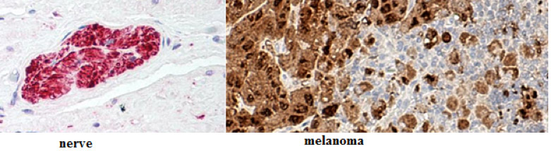
S100 is normally present in cells derived from the neural crest (Neural crest cells are a transient, multipotent, migratory cell population unique to vertebrates that gives rise to a diverse cell lineage including melanocytes, craniofacial cartilage and bone, smooth muscle, peripheral and enteric* neurons and glia), chondrocytes, adipocytes, myoepithelial cells, macrophages, Langerhans cells, dendritic cells, and keratinocytes. It may be present in some breast epithelial cells.
* The enteric nervous system (ENS) or intrinsic nervous system is one of the main divisions of the nervous system and consists of a mesh-like system of neurons that governs the function of the gastrointestinal system.
Several members of the S-100 protein family are useful as markers for certain tumors and epidermal differentiation. It can be found in melanomas, 100% of schwannomas, 100% of neurofibromas (weaker than schwannomas), 50% of malignant peripheral nerve sheath tumors (may be weak and/or focal), paraganglioma stromal cells, histiocytoma and clear cell sarcomas. Further, S100 proteins are markers for inflammatory diseases and can mediate inflammation and act as antimicrobials.
The Histology Lab at The McGowan Institute offers competitive pricing for IHC staining, including “working up” new protocols for Paraffin and Frozen sections. We have S100 already up and running and will waive the work up fee for the month of October! Bring your slides, blocks, frozen or fixed tissue to the histology lab for S100 staining. You are guaranteed beautiful results.
Contact Lori at the McGowan Core Histology Lab and ask about our staining specials. Email perezl@upmc.edu or call 412-624-5265. As always, you will receive the highest quality histology in the quickest turn-around time.
Did you know the more samples you submit to the histology lab the less you pay per sample?
Contact Lori to find out how!
Flow Cytometry Assistance Available at the McGowan Institute
 Flow cytometry is a laser-based, biophysical technology employed in cell counting, cell sorting, biomarker detection, and protein engineering, by suspending cells in a stream of fluid and passing them by an electronic detection apparatus. It allows simultaneous multiparametric analysis of the physical and chemical characteristics of up to thousands of particles per second.
Flow cytometry is routinely used in the diagnosis of health disorders, especially blood cancers, but has many other applications in basic research, clinical practice, and clinical trials. A common variation is to physically sort particles based on their properties, so as to purify populations of interest.
The McGowan Institute for Regenerative Medicine Flow Cytometry Laboratory is ready to help YOU with your research efforts. Do you need to: Purify samples? Characterize complex samples? Define a rare subpopulation? Examine cellular function? Study proliferation or apoptosis? Design a flow experiment and need help? State-of-the-art flow cytometry is available to ALL researchers across the University of Pittsburgh campus. Instrumentation and experienced staff are available to assist users in the understanding, development, and usage of multi-parameter flow cytometry in order to further their research. Contact Lynda Guzik, the Flow Cytometry Lab Manager, for more information: 412-648-8660 or guzilj@upmc.edu.
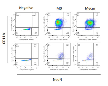
Alessandra Costa, PhD, a postdoctoral fellow in the laboratory of McGowan Institute for Regenerative Medicine deputy director Stephen Badylak, DVM, PhD, MD, recently worked with Ms. Guzik to determine the expression of macrophage and neuronal markers in cells harvested from mouse bone marrow. The Badylak Lab is interested in mechanisms of extracellular matrix (ECM) bioscaffold remodeling and the contribution to constructive and functional tissue replacement. Dr. Costa explains how her experience with the Flow Cytometry Laboratory was instrumental in her investigations:
ECM-based biomaterials have been shown to promote functional tissue reconstruction by inducing stem/progenitor cells recruitment, proliferation, and differentiation, and by influencing the inflammatory response, mainly via modulation of macrophage activity.
The presence of cells expressing both stem and macrophage markers in the area of tissue remodeling in animals treated with ECM-based biomaterials and the ability of these cells to differentiate along various lineages (neurogenic, adipogenic, and osteogenic) is of great interest. Fluorescence-activated cell sorting (FACS) analysis is instrumental in our studies.
We have 6 cell markers in which we are specifically interested, and flow cytometry has allowed us to move forward efficiently and rapidly. We have been able to quantitatively analyze the expression of all the 6 markers simultaneously, even when present in relatively low cell numbers.
Ms. Guzik helped in the creation of a 6-color panel of antibodies and spent many hours helping to optimize a protocol that allows for analysis of surface, cytoplasmic, and nuclear markers. Ms. Guzik’s expertise was influential in moving the project forward.
Figure:M0 and Mecm macrophages before (upper panels) and after (lower panels) a selected treatment were stained for the viability dye eFluor780, CD11b:BUV395, iNos:APC, CD206:BV605, NeuN:biotin and streptavidin 421, β-III-Tubulin:Alexa488, and GFAP:PE. The figure illustrates the analysis of CD11b and NeuN expression by the cell population of interest.
Contact Lynda Guzik, the Flow Cytometry Lab Manager, for more information on how the lab might help YOU: 412-648-8660 or guzilj@upmc.edu.
UPCOMING EVENTS
Save the Date: 9th Symposium on Biologic Scaffolds for Regenerative Medicine
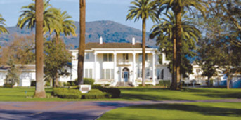 The 9th Symposium on Biologic Scaffolds for Regenerative Medicine will be held at the Silverado Resort in Napa, California on April 28th – 30th, 2016.
This symposium represents an opportunity to advance the use of biologic scaffolds for regenerative medicine and all general surgery applications. Topics will include the basic science of scaffold remodeling from the molecular level through the macroscopic and clinical level. Approximately 50% of the presentations will involve the clinical perspective in the form of formal studies, anecdotal reports, and surgeon reviews. Speakers will provide an objective opinion of the pros and cons of the use of biologic scaffold materials. It is not the intent of this symposium to discuss only the beneficial aspects of biologic scaffolds or particular products, but also to identify problems, develop strategies for solving these problems, and hopefully initiate collaborations among basic scientists, clinicians, and industry in attendance at the meeting.
This symposium is designed to advance the use of biologic scaffolds for regenerative medicine and all general surgery applications via a series of objective presentations describing the potential benefits and risks associated with the use of such materials, factors that affect performance, and the clinical applications that may benefit most from their use. Topics range from the most basic science of scaffold remodeling at the molecular level through the preclinical and clinical level. It is not the intent of this symposium to discuss only the beneficial aspects of biologic scaffolds, but just as importantly to identify the problem areas, develop strategies for solving these problems, and hopefully initiate collaborations among basic scientists and clinicians in attendance at the meeting. Our feedback from previous symposia (this is a bi-annual event) consistently identifies the equal mix of clinicians and basic scientists as the most beneficial and rewarding aspect of the meeting;
Although all topic areas are considered and will be represented, the 2016 symposium will definitely include the following:
- An in depth account of the most recent findings regarding the mechanisms by which biologic scaffolds facilitate constructive remodeling of tissues; including body wall (skeletal muscle), cardiovascular, reconstructive surgical applications such as breast and pelvic floor, and whole organs such as liver, lung and heart.
- Identification and discussion of manufacturing issues such as tissue source, decellularization methods, and sterilization decisions that affect the quality and performance of biologic scaffolds for surgical applications.
- A review of clinical experiences, especially general surgery, orthopedic and trauma related challenges, neurologic applications, gastrointestinal applications, and cardiovascular applications.
- Identification and discussion of the effect of the host innate immune response upon scaffold remodeling and clinical outcome.
List of invited (and accepted) speakers to date: Robert M. Nerem, PhD (Georgia Institute of Technology), Arnold I. Caplan, PhD (Case Western Reserve University), Jeffrey M. Davidson, Ph.D. (Vanderbilt University), Cyrus Ghajar, PhD (Fred Hutchinson Cancer Research Center), Jeffrey A. Hubbell, PhD (Swiss Federal Institute of Technology, EPFL), Kristen Jones, MD (University of Minnesota), C. James Kirkpatrick MD PhD DSc FRCPath (Johannes Gutenberg University), Robert G. Martindale, MD, PhD (Oregon Health & Science University), Charles D. Mills, PhD (BioMedical Consultants), Laura E Niklason PhD, MD (Yale University), Frederick J. Schoen, MD, PhD (Harvard University), Allan S. Stewart, MD (Mount Sinai Hospital NYC), and Nadia Rosenthal, PhD (Monash University, Australia).
SCIENTIFIC ADVANCES
Protein-Hydrogel Combo May Help Heal Heart
 As reported by Prachi Patel of the American Chemical Society’s Chemical and Engineering News, the new strategy to restore damaged cardiac muscle from heart attacks combines a tissue-growing protein with a strengthening gel. Even if a person survives a heart attack, the lack of blood flow damages tissue, which often leads to heart failure. Now McGowan Institute for Regenerative Medicine affiliated faculty member Yadong Wang, PhD, the William Kepler Whiteford Professor in Bioengineering with adjunct positions in Chemical Engineering, Mechanical Engineering and Materials Science, and Surgery at the University of Pittsburgh, and researchers have developed a mixture of a growth protein and a biodegradable gel to prevent scarring and repair injured heart muscle. When injected at the site of a heart attack in pre-clinical trials, the cocktail limited cardiac tissue damage, the team reports.
The new strategy combines two experimental techniques for reviving heart tissue after a heart attack. One approach is to inject a scaffolding material to reinforce the weakened muscle wall. Another is to inject stem cells or growth factors to help repair the damaged tissue. On their own, these individual methods have shown limited success in clinical trials.
Injected growth factors show some promise, says Dr. Wang, but they quickly break down or diffuse out of cardiac tissue.
So Dr. Wang and colleagues devised a special trick to deliver a growth protein trapped within a hydrogel. The gel protects the protein, keeps it at the heart attack site, and supports the damaged walls of the heart right after a heart attack. They used a tissue-signaling protein called Sonic hedgehog that is known to protect cardiac cells and help them grow by inducing the formation of blood vessels.
The researchers mixed it with negatively charged heparin, which is a widely used anticoagulant, and a polycation, which attaches to the oppositely charged heparin molecules to form micrometer-sized liquid droplets.
Next, they combined the droplets with a polyethylene glycol gel. The gel targets the signaling protein to damaged tissue, Dr. Wang says, because it degrades in the presence of an inflammatory enzyme secreted after a heart attack.
If the material shows success in humans, Dr. Wang imagines that doctors could deliver it using a catheter or through a small opening in the chest wall.
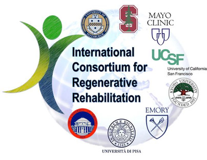
International Consortium for Regenerative Rehabilitation Leadership Council Established
 Medical advances in the field of Regenerative Medicine are accelerating at an unprecedented rate. Biological technologies such as stem cell transplantation, scaffolds, and artificial devices are now being tested in clinical trials throughout the world. With functional outcomes as the ultimate goal of these biological therapies, it is clear that the future of regenerative medicine is tightly intertwined with that of rehabilitation, thus opening up a novel population of patients to clinicians and exciting new areas of investigation for rehabilitation scientists.
For the past 3 years, the University of Pittsburgh in collaboration with the Veteran’s Administration in Palo Alto,California, have administered a Regenerative Rehabilitation Symposium that focuses on fusing regenerative medicine and rehabilitation science, integrating laboratory-based approaches to enhance regeneration with clinically available rehabilitation approaches. The event has drawn over 120 participants in each of the last 3 years and is expected to continue to grow as this novel field expands.
As part of the mission to advance the science of Regenerative Rehabilitation, the Regenerative Rehabilitation Consortium Leadership Council (CLC) was established. Members include:
- Michael Boninger, University of Pittsburgh
- Fabrisia Ambrosio, University of Pittsburgh
- Tom Rando, Stanford University
- Tony Wyss-Coray, Stanford University
- Carmen Terzic, Mayo Clinic
- Chris Evans, Mayo Clinic
- Steve Wolf, Emory University
- Randy Trumbower, Emory University
- George Christ, University of Virginia
- Fred Epstein, University of Virginia
- Linda Noble, University of California at San Francisco
- Kim Topp, University of California at San Francisco
- Tomoki Aoyama, Kyoto University, Kyoto, Japan
- Hiroshi Kuroki, Kyoto University, Kyoto, Japan
- Carmelo Chisari, University of Pisa, Pisa, Italy
It has been determined that the CLC will play a key role in shaping the future of the symposium and the field of Regenerative Rehabilitation by:
- framing the scientific content for the symposium
- becoming leaders in the field and introducing emerging trends, therapies, and other information through the symposium, serving as spokespersons for the field as it emerges
- providing valuable insight and guidance to young investigators, clinicians, therapists, and others interested in pursuing Regenerative Rehabilitation as a career by having first-hand knowledge of potential funding, collaborations, and other opportunities to advance research and clinical activities
- building a new area of science that will have a positive impact on future patient outcomes
The development of the CLC is ongoing. Others are invited to play a role on the Council and in advancing the field of Regenerative Rehabilitation. Interested parties should contact McGowan Institute for Regenerative Medicine executive management team member Patrick Cantini at cantinip@upmc.edu for more information.
Illustration: Regenerative Rehabilitation Consortium.
Children’s Hospital of Pittsburgh Foundation Invests in Cardiovascular Regeneration Research
 Bernhard Kühn, MD, a scientist at the Richard King Mellon Foundation Institute for Pediatric Research at Children’s Hospital of Pittsburgh of UPMC and a McGowan Institute for Regenerative Medicine affiliated faculty member, is being awarded a $200,000 grant from the Children’s Hospital of Pittsburgh Foundation.
The grant is being provided from the Fund for Genomic Discovery, which was raised by the Foundation’s Research and Education Program Committee. Established in 2012, the Research and Education Program Committee promotes the awareness of funding needs and priorities of physician-scientists at Children’s Hospital. Through various fundraising initiatives, the committee seeks to broaden the network of philanthropists, raise money to fund the gaps between government grants, and provide seed funding for new avenues of scientific investigation.
Through hosting two events, combined with additional fundraising efforts, more than $520,000 has been raised for research.
“This funding will allow my team to enter the field of fibrosis research, a new area of investigation for my lab. If successful, this project will provide a broadly applicable molecular-genetic blueprint for the field of cardiovascular development and for developing new drugs to reduce fibrosis in heart disease,” said Dr. Kühn, associate professor of pediatrics at the University of Pittsburgh School of Medicine. “Working directly with me on this research is Dennis Kostka, PhD, an expert in Developmental Biology and Computational & Systems Biology, who will offer his expertise on the computational aspects of the research.”
Dr. Kühn and his team of researchers are focused on cardiomyocytes, the cells of the heart muscle, and discovering ways to make them replicate and proliferate so as to enable the heart to heal itself in cases of heart failure or congenital defects.
“Dr. Kühn is one of the leading researchers in heart regeneration and this funding will give him the opportunity to further explore the growth of heart cells and the advancement of treatments for heart failure,” said McGowan Institute for Regenerative Medicine affiliated faculty member David Perlmutter, MD, physician-in-chief and scientific director, Children’s Hospital, and Distinguished Professor and Vira I. Heinz Endowed Chair, Department of Pediatrics, Pitt School of Medicine.
Studying the Advances in Next-Generation Stem Cell Culture Technologies
 A researcher at West Virginia University (WVU) who is a McGowan Institute for Regenerative Medicine affiliated faculty member is studying ways to advance the next generation of cell culture technologies—the removal of stem cells from an organism and the controlled growth of those cells in an engineering environment—that could treat debilitating diseases.
Stem cells respond to nanoscale features on the cell culture surface. In order to optimize cell culture conditions, Yong Yang, PhD, assistant professor of chemical engineering in the Benjamin M. Statler College of Engineering and Mineral Resources at WVU, is investigating the effect different nanoscale structures have on stem cells.
“Conventional cell culture methods using flat, stiff plastic surfaces do not accurately mimic the characteristics of the microenvironment where cells reside inside the human body,” said Dr. Yang, “which cause cell behaviors on such surfaces to deviate from their live counterparts. For instance, skeletal muscle stem cells lose their regenerative potential rapidly on stiff plastic surfaces, but retain their regenerative capacity on soft hydrogels of physiologically relevant stiffness.
Dr. Yang’s research uses stem cells derived from human bone marrow to test the hypothesis that stem cells can remember the nanoscale surface information from the previous culture environment and that information can influence the regenerative capacity of stem cells. His work is being supported by a grant from the National Science Foundation.
“The nanoscale features critically influence numerous developmental and disease processes, and have a profound influence on stem cell growth and differentiation in cell culture,” Dr. Yang said. “We will engineer a variety of nanotopographies and investigate how stem cells memorize the information from the shape and dimensions of surface features over a certain period of culture time, which eventually has an impact on the regenerative capacity of the stem cell.”
Dr. Anna Balazs’ Manuscript Chosen as an ACS Editors’ Choice from All ACS Journals
 The American Chemical Society (ACS) recently announced that a manuscript by McGowan Institute for Regenerative Medicine affiliated faculty member Anna Balazs, PhD, Distinguished Professor of Chemical Engineering and the Robert v. d. Luft Professor, Department of Chemical & Petroleum Engineering, University of Pittsburgh, published in Langmuir entitled, “Designing Synthetic Microcapsules that Undergo Biomimetic Communication and Autonomous Motion,” has been chosen for an ACS Editors’ Choice from all of the ACS journals.
From the paper’s abstract, inspired by the collective behavior of slime molds and amoebas, Dr. Balazs and colleagues designed synthetic cell-like objects that move and self-organize in response to self-generated chemical gradients, thereby exhibiting autochemotaxis. Using computational modeling, the team specifically focused on microcapsules that encompass a permeable shell and are localized on an adhesive surface in solution. Lacking any internal machinery, these spherical, fluid-filled shells might resemble the earliest protocells. These microcapsules do, however, encase particles that can diffuse through the outer shell and into the surrounding fluid.
Full Text (Designing synthetic microcapsules that undergo biomimetic communication and autonomous motion. Victor V. Yashin, German V. Kolmakov, Henry Shum, and Anna C. Balazs. Langmuir; July 28, 2015.)
Studies into Mitigating Effects of Radiation to Continue
 The University of Pittsburgh Cancer Institute (UPCI) and Pitt’s School of Medicine and Graduate School of Public Health have received a 5-year, $18 million grant from the National Institute of Allergy and Infectious Diseases (NIAID) to continue work developing drugs that could provide protection from radiation in emergencies such as terrorism or reactor meltdowns.
This is the third renewal of the grant for Joel S. Greenberger, M.D., chair of the Department of Radiation Oncology at Pitt, and his team of researchers with the university’s Center for Medical Countermeasures Against Radiation (CMCR). It is one of only four such grants awarded by the NIAID in the U.S.
McGowan Institute for Regenerative Medicine affiliated faculty member Valerian Kagan, PhD, Professor and Vice-Chairman in the Department of Environmental and Occupational Health, is a project leader. McGowan Institute for Regenerative Medicine affiliated faculty members Simon Watkins, PhD, Founder and Director of the Center for Biologic Imaging at the University of Pittsburgh, and Ivet Bahar, PhD, Distinguished Professor, the John K. Vries Chair, and the Founding Chair in the Department of Computational and Systems Biology at the University of Pittsburgh’s School of Medicine, are core leaders.
In its first 10 years, the CMCR has developed and patented two drugs to mitigate the effects of radiation on the body. In this next phase of study, researchers will be looking at ways to administer these drugs individually or in combination using microneedle arrays. Also, they will be working to develop new drugs based on the innovative concept of radiation triggered disease rather than acute injury.
The CMCR consists of four projects and six cores led by a multidisciplinary group of researchers representing Pitt’s School of Medicine and the departments of Critical Care Medicine, Computational Biology, Radiation Oncology, Dermatology, and the Pitt Graduate School of Public Health.
The project leaders are Dr. Kagan; Hülya Bayır, MD; and Jian Yu, PhD. The cores are directed by Michael Epperly, PhD; Dr. Watkins; Peter Wipf, PhD, Dr. Bahar; Yulia Tyurina, PhD; Detcho Stoyanovsky, PhD, and Hong Wang, PhD.
Illustration: Wikipedia.
AWARDS AND RECOGNITION
Dr. William Wagner Named a TERMIS Fellow
TERMIS 2015 Fellows Class
McGowan Institute for Regenerative Medicine Director William Wagner, PhD, Professor of Surgery, Bioengineering, and Chemical Engineering at the University of Pittsburgh, has been named a Fellow of the Tissue Engineering and Regenerative Medicine International Society (TERMIS). Dr. Wagner received this honor, presented by McGowan Institute Deputy Director Stephen Badylak, DVM, PhD, MD, during the 2015 4th TERMIS World Congress recently held in Boston, Massachusetts.
Dr. Wagner’s research interests address medical device biocompatibility and design, tissue engineering, and targeted imaging. Dr. Wagner and his group enjoy working across the spectrum from in vitro to clinical studies.
Dr. Wagner also serves as the Deputy Director of the NSF Engineering Research Center on “Revolutionizing Metallic Biomaterials.” He holds a BS (Johns Hopkins Univ.) and PhD (Univ. of Texas) in Chemical Engineering. Dr. Wagner is the Coordinator for the Cellular and Organ Engineering track for Bioengineering graduate students, and currently teaches in the areas of biomaterials and tissue engineering.
Dr. Wagner is the Founding Editor and Editor-in-Chief of one of the leading biomaterials journals, Acta Biomaterialia, and currently serves on the editorial boards of the Journal of Biomedical Materials Research Part A, Biotechnology and Bioengineering, and the Journal of Tissue Engineering and Regenerative Medicine. Dr. Wagner is also a past president of the American Society for Artificial Internal Organs (ASAIO; 2010-2011) and serves on the Executive Board of the International Federation of Artificial Organs (IFAO). He is a fellow and former vice president of the American Institute for Medical and Biological Engineering (AIMBE; 2000) and has also been elected a fellow of the Biomedical Engineering Society (2007), the International Union of Societies for Biomaterials Science and Engineering (2008), and the American Heart Association (2001). He has served as Chairman for the Gordon Research Conference on Biomaterials: Biocompatibility & Tissue Engineering, as well as for the First World Congress of TERMIS. In 2006 he was selected to the “Scientific American 50,” the magazine’s annual list recognizing leaders in science and technology from the research, business, and policy fields. In 2011 he was awarded the Society for Biomaterials Clemson Award for Applied Research. He has served on numerous NIH and NSF study sections, is a member of the NIH College of Reviewers, and has been a member of external review committees for national and international organizations focused on bioengineering and regenerative medicine. His research has generated numerous patents and patent filings that have resulted in licensing activity, the formation of a company, and University of Pittsburgh Innovator Awards in 2007, 2008, 2009, and 2010.
To accomplish its mission, TERMIS brings together the international community of persons engaged or interested in the field of tissue engineering and regenerative medicine and promotes education and research within the field of tissue engineering and regenerative medicine through regular meetings, publications, and other forms of communication. The Society also serves as an international forum to promote the informed discussion of challenges and therapeutic benefits of the application of tissue engineering and regenerative medicine technologies.
Congratulations, Dr. Wagner!
Badylak Lab High School Student Receives Best Poster Award
 The 2015 UPCI Academy summer session came to a close on Friday, August 7, as the high school students presented their laboratory findings through oral and poster presentations at the Hillman Cancer Center. Congratulations to Zain Mehdi of North Allegheny Senior High School. At the UPCI Summer Academy event, Mr. Mehdi (mentors McGowan Institute for Regenerative Medicine deputy director Stephen Badylak, DVM, PhD, MD, and George Hussey, PhD) received the Best Poster Award among all UPCI summer academy scholars.
The University of Pittsburgh Cancer Institute (UPCI) Academy provides rising high school seniors with 8 weeks of research-focused didactic and experiential learning at seven locations. Scholars work on their own research project in a dedicated research mentor’s laboratory. At the end of the program, scholars are asked to present their projects as an oral presentation and in a poster session. Participating scholars also learn important skills to help prepare them for success in college and in their future careers in science and medicine. McGowan Institute for Regenerative Medicine affiliated faculty member Michael Lotze, MD, is the Director of the UPCI Academy and a Professor of Surgery and Immunology at the UPCI Hillman Cancer Center.
Congratulations, Mr. Mehdi!
Regenerative Medicine Podcast Update
 The Regenerative Medicine Podcasts remain a popular web destination. Informative and entertaining, these are the most recent interviews:
#151 –– Dr. Kelly LaMarco is the Senior Editor and Co-Founder of Science Translational Medicine. Dr. LaMarco discusses the Five Gaps of translational medicine.
Visit www.regenerativemedicinetoday.com to keep abreast of the new interviews.
PICTURE OF THE MONTH
The Picture of the Month is a compliment to the longstanding features Grant of the Month and Publication of the Month. Each of these features highlights the achievements of McGowan affiliated faculty and their trainees. As we have always welcomed suggestions for grants and publications, please also consider submitting images that can highlight your pioneering work.
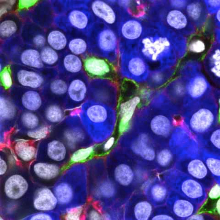
Short description: the zebrafish liver at 76 hours post-fertilization showing Kdrl:GFP (green, endothelial cells) and fabp10a:DsRed (blue, hepatocytes) expression with Hoechst 33342 (grey, nuclei) and phalloidin (red, F-actin) staining. Biliary epithelial cells in the liver strongly express F-actin.
Image by Dr, Juhoon So in Dr. Donghun Shin’s Lab.